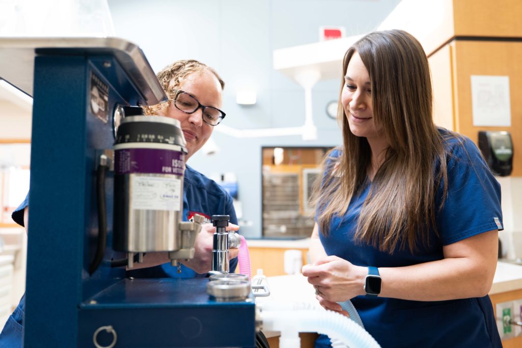
CryoSurgery
Cryosurgery is now being offered at Cleveland Road Animal Hospital for the removal of most skin masses. With cryosurgery, your pet will experience less pain and discomfort than with traditional surgery. Many pets that are unable to go under anesthesia now have the option for this procedure, making it safer and more cost-effective for the removal of most skin masses. Schedule an appointment to see if cryosurgery is right for your pet.
Cruciate Surgery
Cruciate injuries in dogs:
Dogs can tear their cruciate ligament in their knee much like human athletes often do. In fact, the torn cranial cruciate ligament (CCL) is one of the most common knee injuries for dogs. The good news is that we can usually repair this injury here at Cleveland Road Animal Hospital!
What are the signs?
Some dogs will suddenly become lame on the leg and may hold it in the air. Some dogs will have a mild lameness for months that will get better and worse over time. Other dogs will have intermittent mild lameness and then suddenly become very lame.
How is it diagnosed?
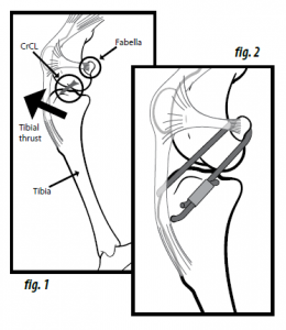
Oftentimes, a veterinarian can diagnose a torn cruciate ligament with a complete physical exam. The knee will feel thickened from the bones shifting back and forth since there is no ligament holding them together, and there will be movement of the knee joint called cranial drawer which is the hallmark of a torn cruciate ligament.
The veterinarian may also do an X-ray. Although you cannot see the ligament on an X-ray, there are classic changes seen on an X-ray such as swelling and edema within the joint. Dogs do not develop arthritis in the knee for no reason, and the most common cause is cruciate tears.
How is a cruciate injury repaired?
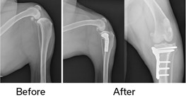
There is not a good way to fix the actual cruciate ligament in dogs, but the most common surgeries to stabilize the knee and alleviate limping are the lateral suture and the TPLO. Today, most veterinary surgeons agree that the TPLO is the best surgery for dogs, especially large breed or more active dogs. TPLO stands for tibial plateau leveling osteotomy.
TPLO Surgery
In this surgery, the surgeon cuts the back part of the tibia with a special circular saw and rotates the piece of bone so the top of the tibia is almost flat. Then the surgeon repairs the bone with a special bone plate to hold it in place while it heals. Once the bone is healed in the “flat” position, the tibia no longer thrusts forward when the dog walks and the knee is stabilized. The plate usually remains on the bone indefinitely, but it is no longer needed for support once the bone heals.
Lateral Suture:
This surgery involves placing a suture, which is usually strong nylon (similar to a strong fishing line), around the outside of the joint to hold the two bones together and prevent them from shifting. The problem with this surgery in large dogs is that there is a lot of force on the nylon and it tends to break and fail. Studies have also shown dogs are in more pain after a lateral suture and develop worse long-term arthritis. Many veterinarians reserve the lateral suture for small breed dogs. Lateral suture surgeries are also less costly, so this may be a better option when trying to stay within a limited budget.
Is knee surgery painful?
Using a saw to make a bone cut is different than a trauma causing the bone to break. The TPLO surgery is usually more comfortable than the lateral suture which feels very tight around the joint. Most dogs are using the leg within a few days post-operatively, and they will often be more athletic after a TPLO than a lateral suture. In both surgeries, the surgery site is treated with a therapy laser to reduce inflammation and pain and patients go home with pain medication. Oftentimes, the surgeon can infuse the surgery site with long-acting bupivacaine that will numb the surgery site for several days after surgery.
How do most dogs do after surgery?
Current studies show that 90-95% of dogs will return to their previous activity after the TPLO. All dogs develop arthritis within a few weeks of the ligament tearing due to inflammation, but once the knee is stabilized, this minimizes arthritis. Dogs will improve up to six months after the surgery as that is how long it takes for them to build muscle strength and for the swelling to improve in the joint. Even with arthritis, most dogs will only have occasional stiff days after strenuous play or long rest but quickly work out of it.
Can there be complications after knee surgery?
The two most common complications are infection and implant failure/fracture of the bone. Infection is the most common with studies showing an infection rate from 8-17%. The most common reason for the infection is that dogs can lick the incision because the E-collar is removed.
If they lick the incision and an infection forms, it can move to cover the plate as there is not a lot of muscle or tissue over the plate. When that happens, it causes a slime coating over the plate that antibiotics cannot penetrate and kill permanently. If an infection forms, the bone usually heals fine, but the implant is often removed to resolve the infection. Fracture/failure of the plate and screws is possible, but this is less common if the dog is kept restricted and there is no jumping or strenuous play.
How long is the recovery for TPLO surgery?
You should confine your dog to controlled leash walks for six to eight weeks post-operatively while the bone heals. They are allowed to walk on the leg, but no jumping, furniture climbing, or running. They should be confined to a crate or very small room while they are healing.
Rehabilitation is recommended to build muscle strength and decrease swelling. At Cleveland Road, our veterinarians will outline some post-op therapy and stretching exercises to do at home and will also recommend seeing the rehabilitation specialists in our area to help with a full return to normal activity.
Patella Luxation Surgery
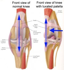
The patella is the medical term for the kneecap. Luxation means that the kneecap dislocates or moves out of its normal position. A medial patella luxation (MPL) occurs when the kneecap moves out of its normal position toward the inside of the leg. Less common is the lateral patella luxation, which is when the kneecap moves toward the outside of the leg.
What does Medial Patella Luxation do?
The patella normally sits in a deep groove located at the end of the femur (thigh bone). It glides smoothly over the cartilage within the groove during knee movement. In patients with patella luxation, this groove is usually too shallow – allowing the kneecap to “pop out.” Luxation can worsen and increase in frequency over time due to constant dislocation. The great majority of patella luxations develop as pets grow. In many breeds, it is considered a genetic or inherited trait. Most patients will have patella luxation by the time they reach maturity, but may not develop signs of problems until later in life.
How can a MPL be fixed?
There are four steps to repairing a luxated patella. Each patient is different. Your pet may require some or all of the steps for correction. The surgeon will discuss their plan for your pet and the specific steps required.
- Medial desmotomy: This step loosens the tissue on the side of the knee that is too tight and contributes to pulling the kneecap out of place.
- Lateral imbrication: This step tightens the loose tissue on the side of the knee to help keep the kneecap in the groove.
- Deepening of the shallow groove is achieved by surgically reshaping the width and depth of the groove using special instruments. This allows the patella to rest deeply within the groove and move appropriately during knee movement.
- Tibial tuberosity transposition is achieved by separating the bone to which the patella attaches from its present position. It is then moved to a new position and maintained in its new placement with bone pins. The new location keeps the tendon in proper place to prevent luxation.
Does surgery work?
At Cleveland Road, we define success as a dramatic improvement in limb function, permanent relocation of the patella, and return to normal or near-normal activity. This is expected in >90% of patients.
The complication rate for these procedures is low. Infections uncommonly develop and when they do, usually resolve completely with proper treatment. Rare complications include pin breakage and migration. Anesthesia also carries a small risk of complications, however, we use the same drugs and monitoring equipment that are used in humans, and anesthetic complications are rare. If your pet has any medical conditions such as heart murmurs, kidney disease, breathing difficulties, or liver disease, please be sure to discuss these preexisting conditions with us so that appropriate steps can be taken to ensure safety with anesthesia for your pet.
What happens after surgery?
The majority of postoperative care involves the restriction of your dog’s activity. No off-leash activities are allowed. Inside the house, your pet should avoid flights of stairs and slippery floors. No running, jumping, or playing is allowed for the first six weeks after surgery. When your dog is not under your direct control, he/she should be confined to a small room, cage, or crate.
Explore Our Complete List of Veterinary Services in Wooster, OH
What's Next
Call us or schedule an appointment online.
Meet with a doctor for an initial exam.
Put a plan together for your pet.